

What Color Is Cancer On Scan Stomach
Abdominal CT scan
Computed tomography scan - abdomen; CT browse - belly; CT abdomen and pelvis
An abdominal CT scan is an imaging method. This exam uses x-rays to create cantankerous-sectional pictures of the belly area. CT stands for computed tomography.
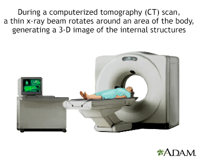
CT stands for computerized tomography. In this procedure, a thin X-ray beam is rotated around the area of the trunk to be visualized. Using very complicated mathematical processes chosen algorithms, the computer is able to generate a 3-D image of a section through the trunk. CT scans are very detailed and provide excellent information for the physician.
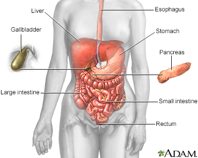
The esophagus, stomach, large and minor intestine, aided past the liver, gallbladder and pancreas convert the nutritive components of food into energy and break downward the not-nutritive components into waste to be excreted.
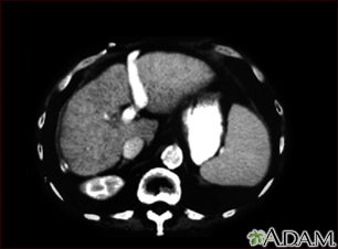
A CT scan of the upper abdomen showing cirrhosis of the liver.
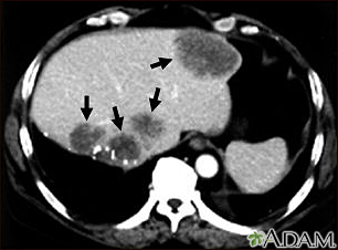
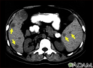
A CT scan of the upper abdomen showing multiple metastasis (cancer that has spread) in the liver of a patient with carcinoma of the large bowel. Note the dark areas in the liver (left side and middle of flick).
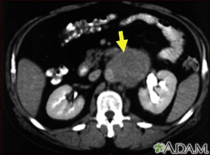
A CT scan of the eye abdomen showing a large tumor mass due to metastasis (spreading cancer) in abdominal lymph nodes.
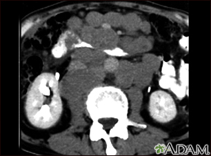
This intestinal CT browse shows tumor masses (malignant lymphomas) in the area behind the peritoneal cavity (retroperitoneal infinite).
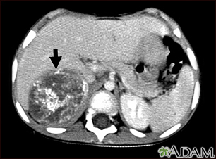
This CT scan of the upper abdomen shows a large tumor (neuroblastoma) on the person's right side (lower left side of picture show). The tumor is behind the liver and is pushing the liver forward and may take perhaps spread into the liver tissue.
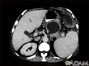
A CT scan of the upper belly showing a large cyst in the pancreas (cystic adenoma of the pancreas) seen on the upper right side of the pic.
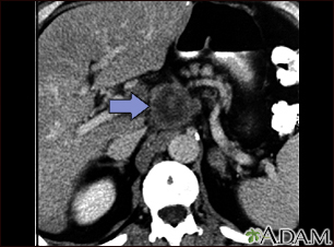
A CT browse of the upper abdomen showing a tumor (pancreas carcinoma) in the head of the pancreas, seen hither in the middle of the movie.
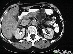
A CT scan of the upper abdomen showing a pseudocyst in the corpus, or tail, of the pancreas.
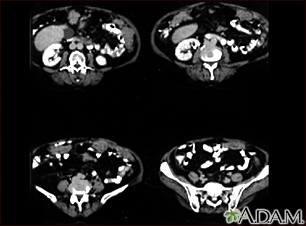
A CT scan serial of the lower abdomen showing ovarian cancer that has metastasized (spread) to the peritoneum.
This CT scan of the upper abdomen shows multiple tumors in the liver and spleen that accept spread (metastasized) from an original intestinal cancer (carcinoma).
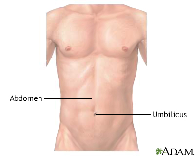
The belly is the area of the torso between the chest and pelvis. Some of the big internal organs comprised in this surface area are the liver, tum, kidneys, and intestines.
How the Test is Performed
Yous will prevarication on a narrow tabular array that slides into the centre of the CT scanner. Most often, y'all will lie on your dorsum with your artillery raised above your head.
Once you are inside the scanner, the motorcar's x-ray beam rotates around you lot. Modern spiral scanners can perform the exam without stopping.
A computer creates split up images of the belly area. These are called slices. These images can be stored, viewed on a monitor, or printed on film. Three-dimensional models of the belly area tin can be made by stacking the slices together.
Y'all must be yet during the exam, considering movement causes blurred images. You may be told to agree your breath for short periods of time.
In many cases, an intestinal CT is done with a pelvis CT.
The scan should accept less than thirty minutes.
How to Prepare for the Examination
You demand to take a special dye, called contrast, put into your body before some exams. Dissimilarity helps certain areas prove up ameliorate on the x-rays. Contrast can be administered in various ways. Such as:
- Contrast tin be given through a vein (IV) in your paw or forearm. If contrast is used, you may also be asked non to swallow or drink anything for 4 to six hours before the exam.
- You may take to drink the contrast before the exam. When y'all potable it will depend on the type of examination being washed. Dissimilarity has a chalky taste, although some are flavored and then they gustation a little improve. The contrast you lot drink will laissez passer out of your trunk through your stools and is harmless.
Let your health care provider know if you have ever had a reaction to contrast. You may need to take medicines before the test to safely receive this substance.
Earlier receiving the contrast, tell your provider if yous take the diabetes medicine metformin. People taking this medicine may have to end taking it for a while earlier the test.
Permit your provider know if you have whatever kidney bug. The Iv dissimilarity can worsen kidney function.
Too much weight can damage the scanner. Detect out if the CT machine has a weight limit if y'all counterbalance more than 300 pounds (135 kg).
You will need to have off your jewelry and article of clothing a infirmary gown during the study.
How the Exam will Feel
Lying on the hard table may be a little bit uncomfortable.
If you have contrast through a vein (IV), you lot may have:
- Slight called-for sensation
- Metallic gustatory modality in the mouth
- Warm flushing of the body
These feelings are normal and get away within a few seconds.
Why the Exam is Performed
An abdominal CT browse makes detailed pictures of the structures within your belly very rapidly.
This examination may exist used to await for:
- Crusade of claret in the urine
- Crusade of intestinal pain or swelling
- Cause of abnormal claret exam results such as liver or kidney problems
- Hernia
- Cause of a fever
- Masses and tumors, including cancer
- Infections or injury
- Kidney stones
- Appendicitis
What Abnormal Results Mean
The abdominal CT scan may bear witness some cancers, including:
- Cancer of the renal pelvis or ureter
- Colon cancer
- Hepatocellular carcinoma
- Lymphoma
- Melanoma
- Ovarian cancer
- Pancreatic cancer
- Pheochromocytoma
- Renal prison cell carcinoma (kidney cancer)
- Spread of cancers that began outside the belly
The abdominal CT browse may prove problems with the gallbladder, liver, or pancreas, including:
- Acute cholecystitis
- Alcoholic liver illness
- Cholelithiasis
- Pancreatic abscess
- Pancreatic pseudocyst
- Pancreatitis
- Blockage of bile ducts
The intestinal CT scan may reveal the post-obit kidney problems:
- Blockage of the kidneys
- Hydronephrosis (kidney swelling from the backflow of urine)
- Kidney infection
- Kidney stones
- Kidney or ureter damage
- Polycystic kidney disease
Abnormal results may also be due to:
- Abdominal aortic aneurysm
- Abscesses
- Appendicitis
- Bowel wall thickening
- Crohn disease
- Renal artery stenosis
- Renal vein thrombosis
Risks
Risks of CT scans include:
- Allergy to contrast dye
- Exposure to radiation
- Damage to kidney role from dissimilarity dye
CT scans betrayal you to more radiation than regular x-rays. Many 10-rays or CT scans over time may increase your risk for cancer. All the same, the risk from any one scan is small. Near mod scanners are able to reduce the radiation exposure. Talk to your provider about this risk and the benefit of the test for getting a correct diagnosis of your medical problem.
Some people take allergies to contrast dye. Let your provider know if you have ever had an allergic reaction to injected contrast dye.
The nigh common type of dissimilarity given into a vein contains iodine. If y'all have an iodine allergy, you may have nausea or vomiting, sneezing, itching, or hives if y'all get this blazon of contrast. If you must be given such contrast, your provider may give you antihistamines (such every bit Benadryl) or steroids earlier the test.
Your kidneys help remove Iv dye from the body. Y'all may need extra fluids after the test to help affluent the iodine out of your trunk if you lot have kidney disease or diabetes.
Rarely, the dye may crusade a life-threatening allergic response. Tell the scanner operator correct abroad if yous have any trouble breathing during the test. Scanners come up with an intercom and speakers, so the operator tin hear you lot at all times.
References
Al Sarraf AA, McLaughlin PD, Maher MM. Electric current status of imaging of the gastrointestinal tract. In: Adam A, Dixon AK, Gillard JH, Schaefer-Prokop CM, eds. Grainger & Allison's Diagnostic Radiology: A Textbook of Medical Imaging. 7th ed. Philadelphia, PA: Elsevier; 2021:chap 18.
Levin MS, Gore RM. Diagnostic imaging procedures in gastroenterology. In: Goldman L, Schafer AI, eds. Goldman-Cecil Medicine. 26th ed. Philadelphia, PA: Elsevier; 2020:chap 124.
Smith KA. Abdominal hurting. In: Walls RM, Hockberger RS, Gausche-Loma M, eds. Rosen's Emergency Medicine: Concepts and Clinical Practice. 9th ed. Philadelphia, PA: Elsevier; 2018:chap 24.
Version Info
Last reviewed on: 7/three/2020
Reviewed by: Jason Levy, MD, Northside Radiology Associates, Atlanta, GA. As well reviewed by David Zieve, Physician, MHA, Medical Director, Brenda Conaway, Editorial Director, and the A.D.A.M. Editorial team.


What Color Is Cancer On Scan Stomach,
Source: https://www.mountsinai.org/health-library/tests/abdominal-ct-scan
Posted by: blackwithander.blogspot.com
0 Response to "What Color Is Cancer On Scan Stomach"
Post a Comment